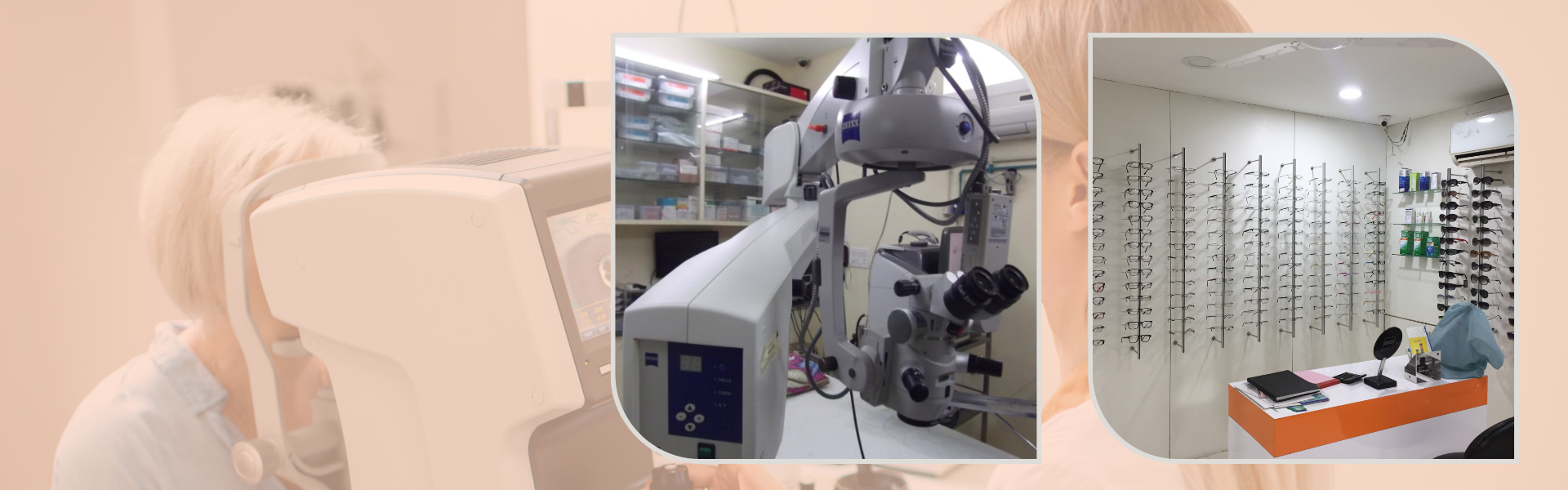
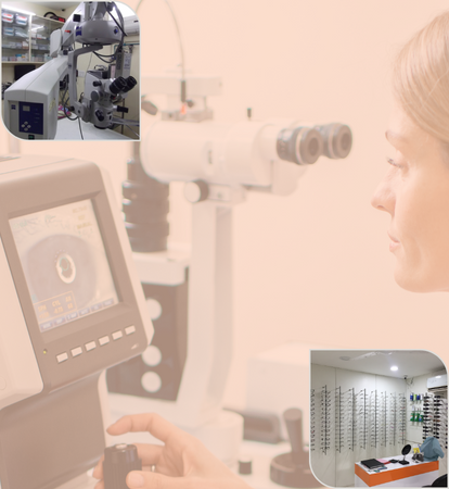
Total eye checkup
This section will help you in understanding some of the terminology your Doctor uses at the time of the examination of your eye. It also details the various tests and surgeries done on eye depending on the symptoms.
Complaints:
- Loss of Vision and Blurred Vision
- Double Vision
- Redness of the Eye
- Stickiness of the Eye
- Watering of the Eye
- White Reflex in the Eye
- Abnormal Looking Eye
- Dropping Eye Lid
- Squinting of the Eye
Ophthalmology Terms:
- Vision Testing
- Refraction
- Amsler Grid Testing
- Dilation
- Slit Lamp Examination
- Applanation Tonometry
- Ophthalmoscopy
Special Tests for Glaucoma:
- Optic Disc Photography
Special Tests for Neurophthalmology:
- Contrast Sensitivity Testing
- Color Vision Testing
Special Tests for Retinal Diseases and Uveitis
- Fundus Photography
- Fundus Fluorescein Angiography (FFA)
- Low Vision Aid Testing
Complaints:
Loss of Vision and Blurred Vision
- Vision can be defective to a variable degree. It may be easy to detect gross decrease in vision but it may be more difficult to detect subtle degree of loss of vision. It is very easy to miss gross loss of vision in one eye when the other eye is healthy unless one consciously tests each eye separately. It is a good practice to test each eye separately at regular intervals using any fine reading material such as newspaper.
Double Vision
- Normally the image formed by the two eyes is coordinated into a single image by the brain. Two distinct images are seen once this coordination is disturbed due to various diseases involving the muscles of the eye and the nerves that control the same. Multiple images often are an early symptom of cataract.
Redness of the Eye
- Visible redness of the one or both eyes is a common symptom pertaining to several varieties of diseases. One should not mistake every red eye as having viral conjunctivitis (So called Madras eye). Hence do not self medicate and delay seeking medical advice if you have a red eye. It could be something serious.
Stickiness of the Eye
- A common symptom of infection in the eye is stickiness of the eyelids due to discharge. This infection could be purely external or could be more serious. Persistent stickiness of the eye lashes needs early evaluation.
Watering of the Eye
- Watering could be the result of mal alignment of the eyelids or eyelashes or a blockade of tear ducts that normally drain the tear fluid into the nose. Presence of tearing in newborn babies can indicate lack of patency of the tear ducts and may need attention.
White Reflex in the Eye
- Normally the center of the eye gives a black reflex due to the pupil. A white reflex can be due to opacification of the normally transparent cornea, the lens (cataract) or due to an abnormal growth of tissue behind the lens. A white reflex in a child can potentially be dangerous and should not be ignored.
Abnormal Looking Eye
- Abnormal look of the eye could be due to prominence of the eye, or could be the result of defects involving the eyelids. Prominence of the eye could be due to large eyeballs or due to protrusion of normal sized eye by abnormal growth behind the eye. Any change in appearance of the eye should be investigated. Previous photographs could be useful in comparing especially when one is not certain about the time of onset of the abnormal look of the eye.
Drooping Eye Lid
- Drooping of the upper eyelid could be present at birth or could occur later. If the defect has occurred later in life one should note the frequency of the occurrence and in what part of the day it is more prominent. These observations can help the doctor make important decisions.
Squinting of the Eye
- Squinting indicates the misalignment of the eyes. In children, this can potentially lead to reduction of vision in the squinting eye due to disuse (lazy eye). When in doubt, taking photographs with flash can help identify the squint in the photographs. This is especially useful to the doctor, in case of children who refuse to cooperate with the doctor for adequate examination.
Ophthalmology Terms:
Vision Testing
- Vision testing involves making a person read standard sized letters at a specified distance. The doctors record the vision as a fraction e.g. 6/6 etc. The top number denotes the distance (in feet) at which the patient has been able to read the particular sized letter while the bottom number indicates the distance at which a normal person is expected to read the same letter. Near vision is tested separately in good illumination using special test charts held at normal reading distance. The testing is done with each eye separately. The doctors often test the vision using a pinhole. This gives an estimate of improvement possible with glasses. The patient in place of glasses cannot use the pinhole.
Refraction
- This is an important test that is done by the ophthalmologist or more often by the optometrist. The eye is like a camera. The light rays are focused on to the light sensitive film in the back of the eye called the retina. This focusing is made possible by the cornea (a clear watch glass like structure in the front of the eye) and by the lens in the eye (similar to the lens of a camera). Refraction is done usually in the normal state. On occasion (especially in children) it may have to be done using special eye drops (cycloplegics). In this situation one may have to retest the power of the required glasses 2-3 days after the testing with the use of drops.
- Refraction involves two parts. The first part is objective where in the refractionist estimates the power needed by using a test called retinoscopy. This test can also be done with a machine called the automatic refractometer (so called computer testing). However one still needs to do the all-important subjective testing (i.e. testing the response of the patient with different powered glasses) before prescribing the glasses. Hence do not be misled by the so-called computer testing.
Amsler Grid Testing
- This test is done in selected group of patients depending upon their symptoms. The test involves looking at a chart that has a grid drawn with a central dot. The test is done using the near vision glasses (if one is using the same). The test permits the evaluation of function of the central 20 degrees of the retina.
- In this test, the patient is asked to look at the central dot and tell whether all the corners of the chart are seen. All the lines are seen straight and not crooked There are any areas of gray patches where the lines are not seen. Whether the central part of the chart or the peripheral part of the chart is clear. The Amsler's chart is very useful as a home monitoring device. If any defect is noted, immediate ophthalmologic examination is warranted.
Dilation
- One of the most common procedures that is done in an eye specialist's office is dilatation. The pupils of the eye constrict or dilate depending upon the light that thrown at the eye. For examining the back part of the eye (fundus), the doctor uses an instrument called ophthalmoscope. To get a good view of the back of the eye, one needs to dilate the pupils. This permits more light to enter the eye and gives a better image of the fundus. To keep the pupils dilated despite the intense light, one needs to dilate the pupils. There are various types of dilating drops available. The faster acting ones may dilate the pupil in 15-20 minutes time.
- Other variety of drops may take up to 30-45 minutes for good dilatation. The effect of dilatation usually lasts up to 6 hours. Some of them may retain the effect for 24 hours. Usually the drops used for routine eye examination do not have long lasting effect. A patient is expected to have glare in the sun light while still under the effect of the drops. Hence driving may become difficult. If you had similar dilatation in the past and have been noted to be allergic to any one of them, please inform the same to your doctor.
Slit Lamp Examination
- Slit lamp is an instrument that has an in built microscope and a bright illumination system. The special arrangement of the light and the microscope allows the doctor to view the eye in great detail under high magnification. The front part of the eye is examined without any other aids while the back part of the eye (fundus) is examined with help of special lenses held in front of the eyeball.
Applanation Tonometry
- In this a small prism mounted on the slit lamp is used to contact the eyeball and measure the pressure. This modality of testing is more accurate and is the standard today.
Ophthalmoscopy
- This is a very important step in the total examination of the eyes. The visible portion of the eye is easily examined by the slit lamp examination. The back portion of the eye can only be examined by using the ophthalmoscope. This step usually needs dilatation of the pupils. This test involves throwing bright light into the eye and examining the image of the back of the eye using special lenses. For Indirect opththalmoscopy, the patient has to be in the reclining position for proper examination. Sometimes the slit lamp may be used for detailed evaluation of the areas of the back of the eye such as macula, optic disc etc.
Special Tests for Glaucoma:
Optic Disc Photography
- Optic disc is the only part of the optic nerve visible to the eye doctor in the back of the eye. The appearance of the disc gives valuable information to diagnose and treat conditions such as glaucoma. It is important to be able to compare the appearance of the disc over a period time in cases of chronic glaucoma. This is made possible by several techniques- one of which is the photography of the disc using the fundus camera.
Special Tests for Neurophthalmology:
Contrast Sensitivity Testing
- Certain disease of the retina and optic nerve leave behind subtle defects of sensitivity. A patient is very symptomatic of these deficiencies but the commonly performed tests like the vision testing do not reveal the true extent of the defect. Measurement of contrast sensitivity enables one to understand these subtle defects in the visual function. This test involves identification of patterns of gray on gray background.
Color Vision Testing
- Color vision is an important component of human vision. Defects in this can be by birth or due to any acquired diseases. The testing is done using one of the two methods.
Special Tests for Retinal Diseases and Uveitis
Fundus Photography
- Fundus photography permits documentation of the structures of the eye. This documentation may be important to compare with other investigations such as fluorescein angiography as well as for follow up. Fundus photography of the optic disc is important in the management of glaucoma.
Fundus Fluorescein Angiography (FFA)
- This is an important test to evaluate a variety of retinal disease such as diabetic retinopathy. This is one of the commonest tests performed for retinal diseases. The test involves injecting a dye called sodium fluorescein into the blood stream and taking photographs of the retina using special filters. The test is important to stage the disease as well as to guide treatment with laser photocoagulation. Present generation digital cameras permit manipulation of the pictures and for instant viewing without need for development of the film etc.
Low Vision Aid Testing
- There are certain diseases that may lead to permanent partial loss of vision. These patients can be sometimes helped to some extent by using special aids called low vision aids. There are a variety of these available and most of them are fine tuned for a specific function. Most of them have been made to enable reading fine print. It is important that the patient should be motivated to use them. They are used at a closer range than normal working distance and hence one needs to get used to the same. Computers and closed circuit television are also useful as low vision aids. One has to test different varieties before choosing what is appropriate for them.
Cataract surgery (phaco surgery /MICS)
Lorem ipsum dolor sit amet consectetur, adipisicing elit. Vero dolore voluptas fugit cupiditate nihil aperiam quo corporis excepturi incidunt praesentium! Quaerat alias corrupti neque, nihil consequuntur quam dolorum numquam? Voluptates.
Lasik surgery
Lorem ipsum dolor sit amet consectetur, adipisicing elit. Vero dolore voluptas fugit cupiditate nihil aperiam quo corporis excepturi incidunt praesentium! Quaerat alias corrupti neque, nihil consequuntur quam dolorum numquam? Voluptates.
Squint surgery
Lorem ipsum dolor sit amet consectetur, adipisicing elit. Vero dolore voluptas fugit cupiditate nihil aperiam quo corporis excepturi incidunt praesentium! Quaerat alias corrupti neque, nihil consequuntur quam dolorum numquam? Voluptates.
Glaucoma treatment
Lorem ipsum dolor sit amet consectetur, adipisicing elit. Vero dolore voluptas fugit cupiditate nihil aperiam quo corporis excepturi incidunt praesentium! Quaerat alias corrupti neque, nihil consequuntur quam dolorum numquam? Voluptates.
Pterygium surgery
Lorem ipsum dolor sit amet consectetur, adipisicing elit. Vero dolore voluptas fugit cupiditate nihil aperiam quo corporis excepturi incidunt praesentium! Quaerat alias corrupti neque, nihil consequuntur quam dolorum numquam? Voluptates.
Contact lens clinic
Lorem ipsum dolor sit amet consectetur, adipisicing elit. Vero dolore voluptas fugit cupiditate nihil aperiam quo corporis excepturi incidunt praesentium! Quaerat alias corrupti neque, nihil consequuntur quam dolorum numquam? Voluptates.